1aHumans reproduce ________.
AnswerSexually.
Learn the complete human reproductive sequence — male and female reproductive systems, gametogenesis, menstrual cycle, fertilisation, implantation, pregnancy, parturition and lactation — in a topic-wise board-exam friendly format.
Read the whole reproductive sequence before studying each system in detail.
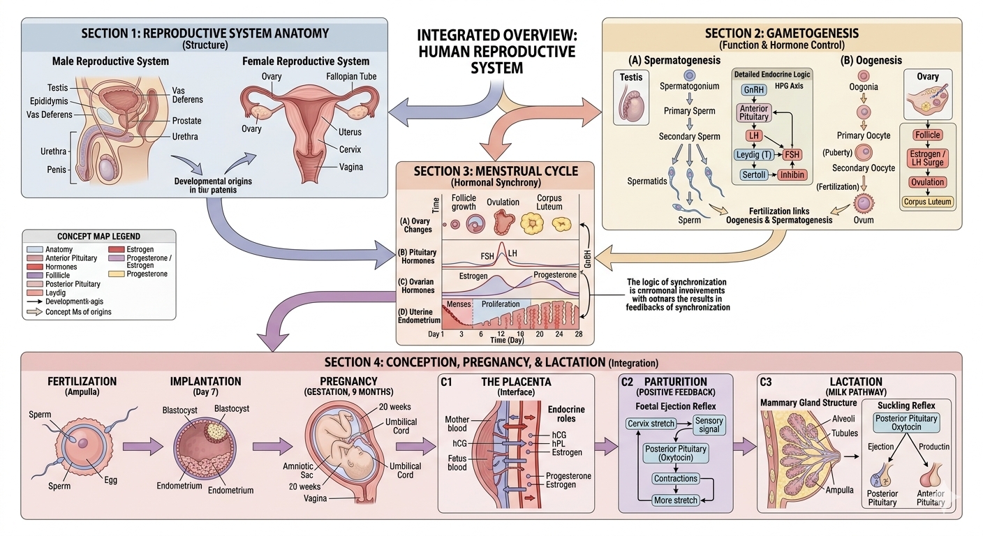
Human reproductive events occur after puberty and include formation of gametes (gametogenesis), transfer of sperm into the female genital tract (insemination), fusion of male and female gametes (fertilisation), zygote formation, cleavage, blastocyst formation and implantation, embryonic development during gestation, delivery of the baby by parturition and feeding of the newborn by lactation.
The chapter moves from gamete formation → fertilisation → development → birth → lactation.
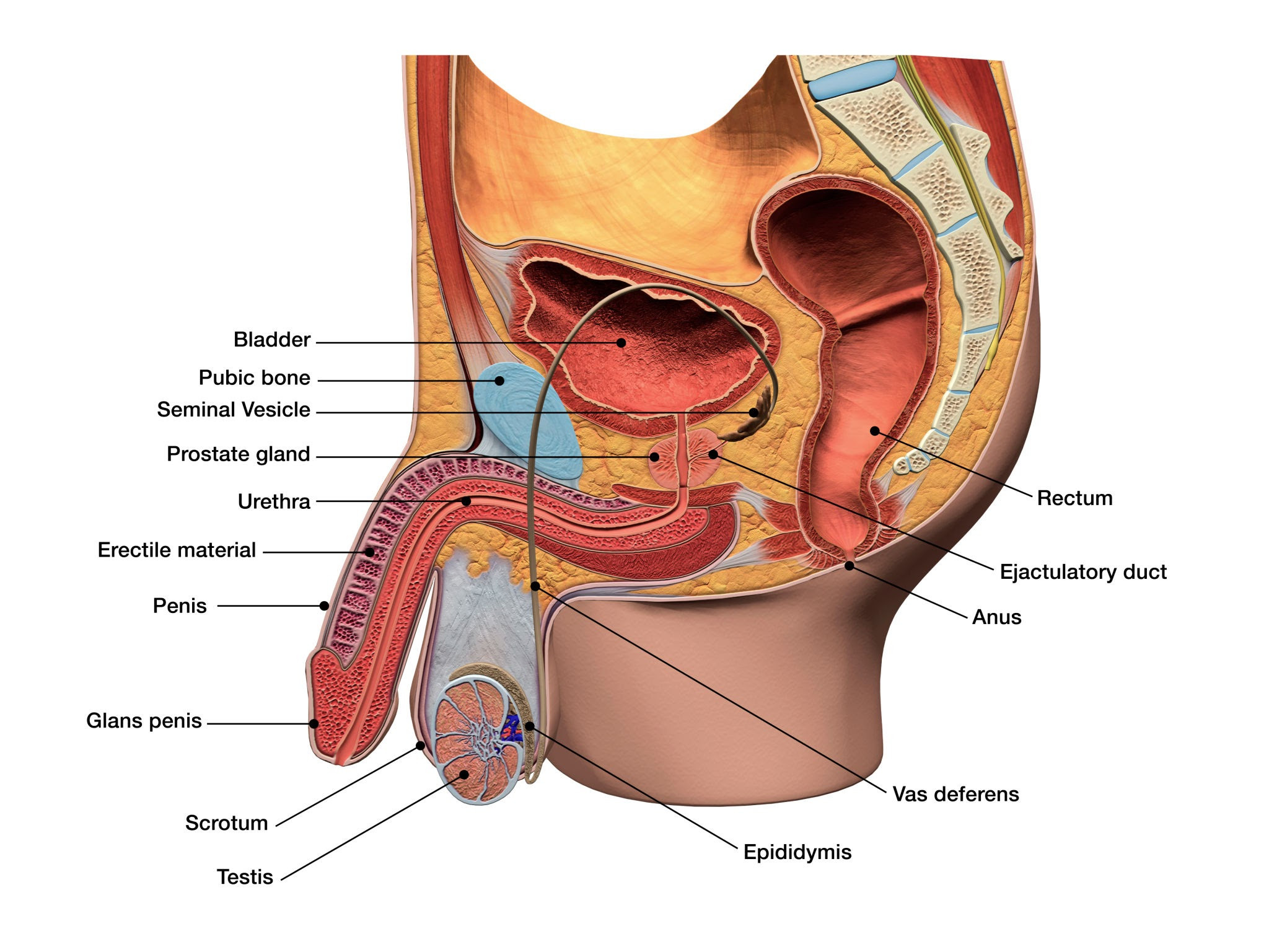
Structures involved in sperm formation, transport and insemination.
The male reproductive system is located in the pelvic region. It includes a pair of testes along with accessory ducts, accessory glands and external genitalia.
The testes are situated outside the abdominal cavity in the scrotum. The scrotum maintains the temperature of the testes about 2–2.5°C lower than normal internal body temperature, which is necessary for spermatogenesis. In adults, each testis is oval, about 4–5 cm long and 2–3 cm wide. Each testis has about 250 testicular lobules, and each lobule contains one to three highly coiled seminiferous tubules.
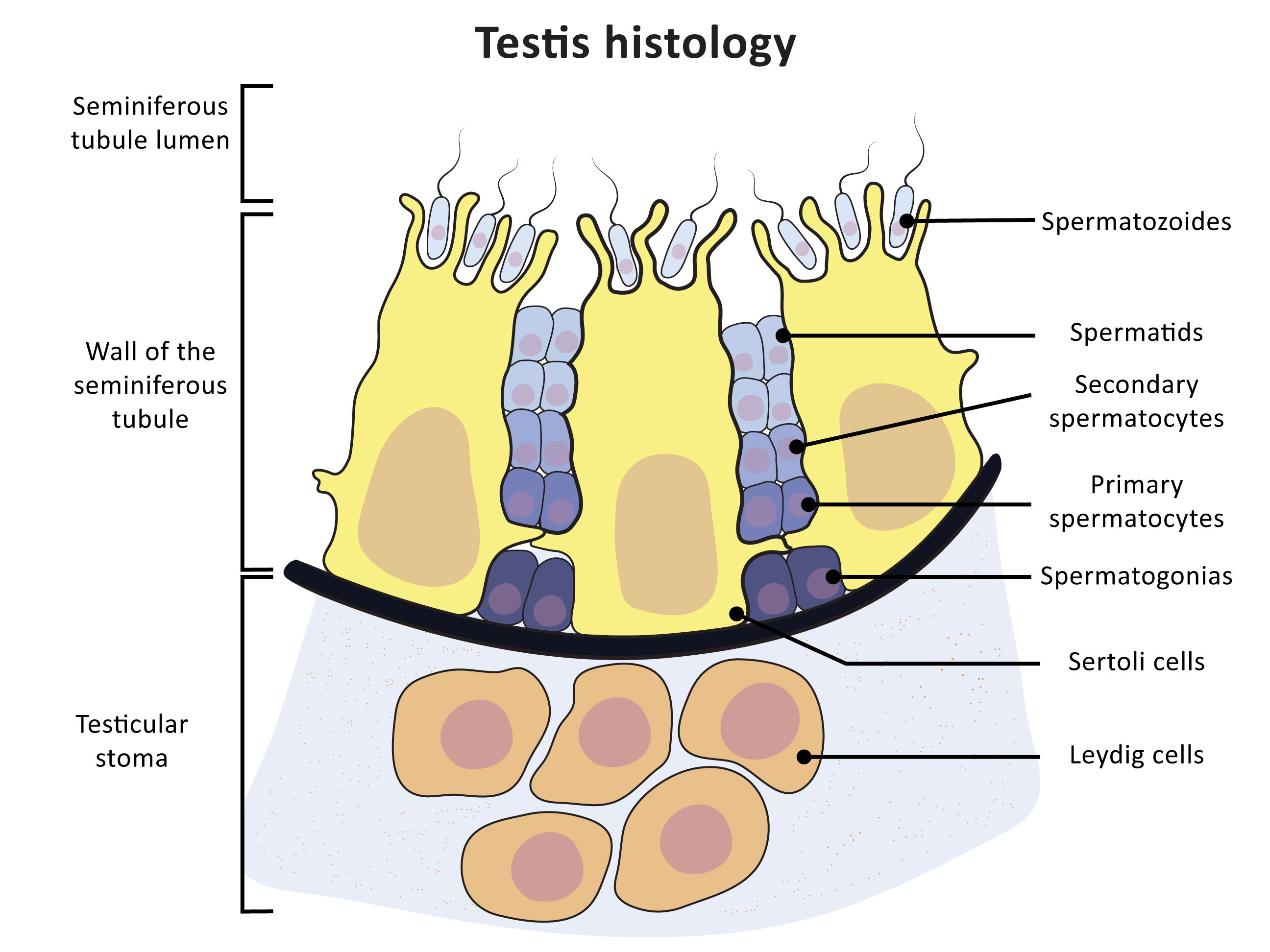
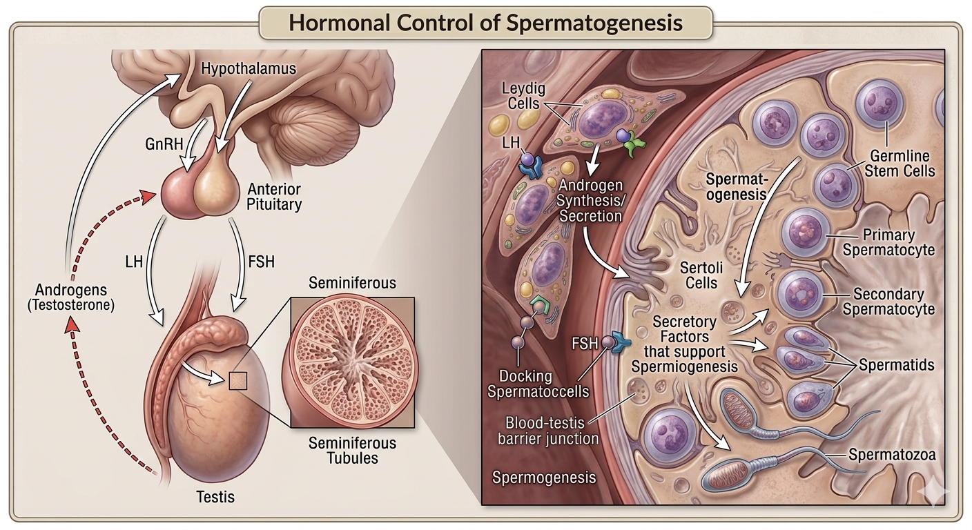
Seminiferous tubules are the sites where sperm are produced. Their inner lining contains spermatogonia and Sertoli cells. Spermatogonia undergo meiotic divisions leading to sperm formation, while Sertoli cells provide nutrition to developing germ cells. Interstitial spaces outside the tubules contain blood vessels and Leydig cells, which synthesise and secrete androgens.
Accessory ducts include rete testis, vasa efferentia, epididymis and vas deferens. They store and transport sperm. Accessory glands include paired seminal vesicles, a prostate and paired bulbourethral glands. Their secretions form seminal plasma, rich in fructose, calcium and certain enzymes; bulbourethral secretions also aid lubrication.
The penis is the male external genitalia. Special tissue helps erection for insemination. The enlarged end is the glans penis, covered by foreskin. The urethra extends through the penis to the urethral meatus.
Structures that support ovulation, fertilisation, pregnancy, birth and child care.
The female reproductive system consists of a pair of ovaries, a pair of oviducts, the uterus, cervix, vagina, external genitalia and a pair of mammary glands.
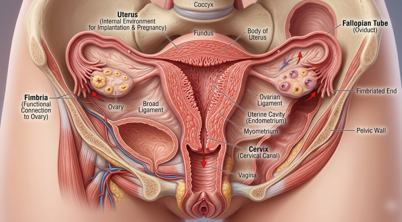
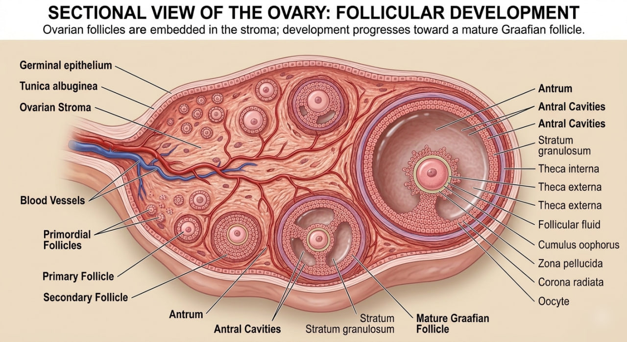
Ovaries are the primary female sex organs. They produce the female gamete (ovum) and several steroid hormones. Each ovary is about 2–4 cm long and is connected to the pelvic wall and uterus by ligaments. The ovary is covered by thin epithelium and its stroma is divided into a peripheral cortex and inner medulla.
Each fallopian tube is about 10–12 cm long. The part nearer the ovary is the funnel-shaped infundibulum with finger-like fimbriae. It leads to the wider ampulla. The isthmus is the last, narrower part and joins the uterus. Fimbriae help in collection of the ovum after ovulation.
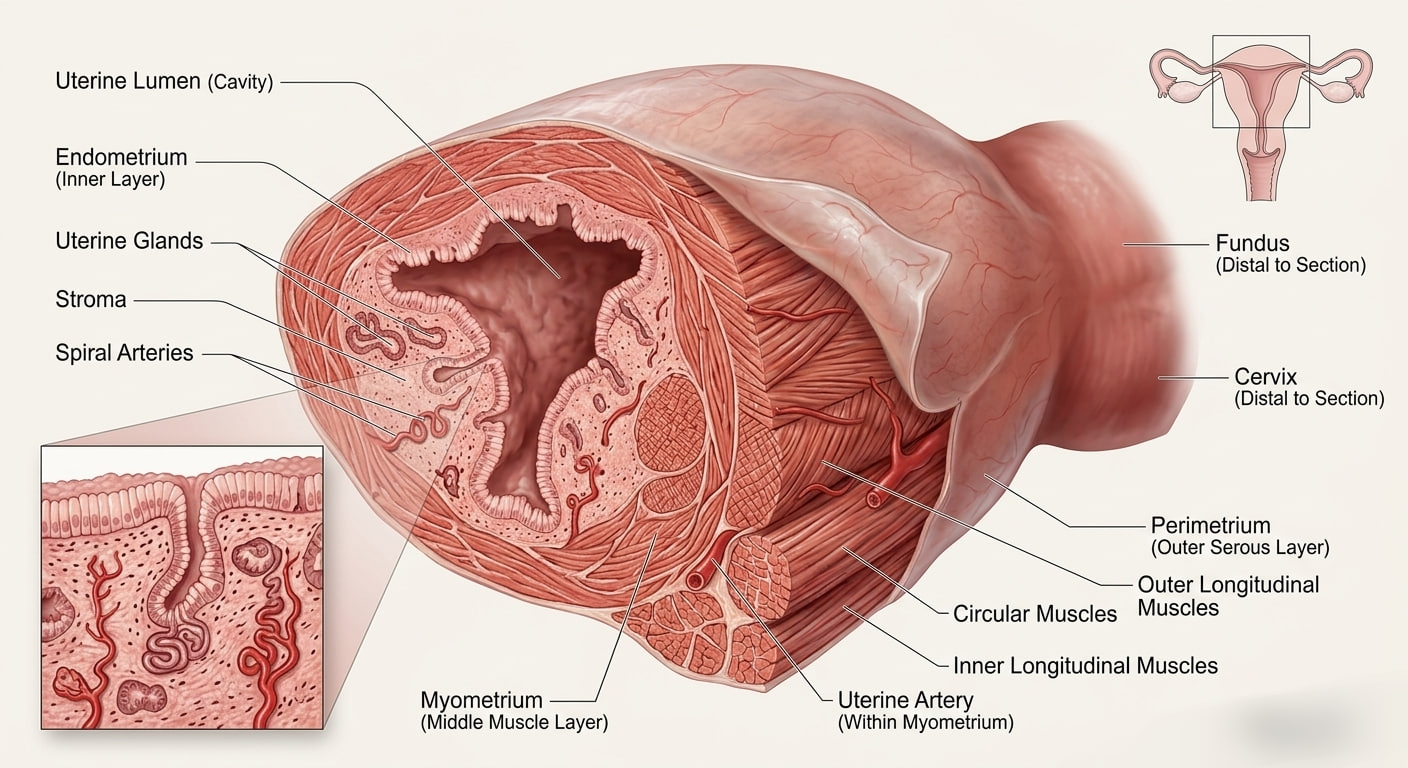
The uterus is a single womb shaped like an inverted pear. The uterus opens into the vagina through the narrow cervix. The cervical canal together with the vagina forms the birth canal. The uterine wall has three layers: outer perimetrium, middle myometrium and inner endometrium. Endometrium undergoes cyclical changes during the menstrual cycle; myometrium contracts strongly during delivery.
The source lists mons pubis, labia majora, labia minora, hymen and clitoris. The source also states that presence or absence of hymen is not a reliable indicator of virginity or sexual experience.
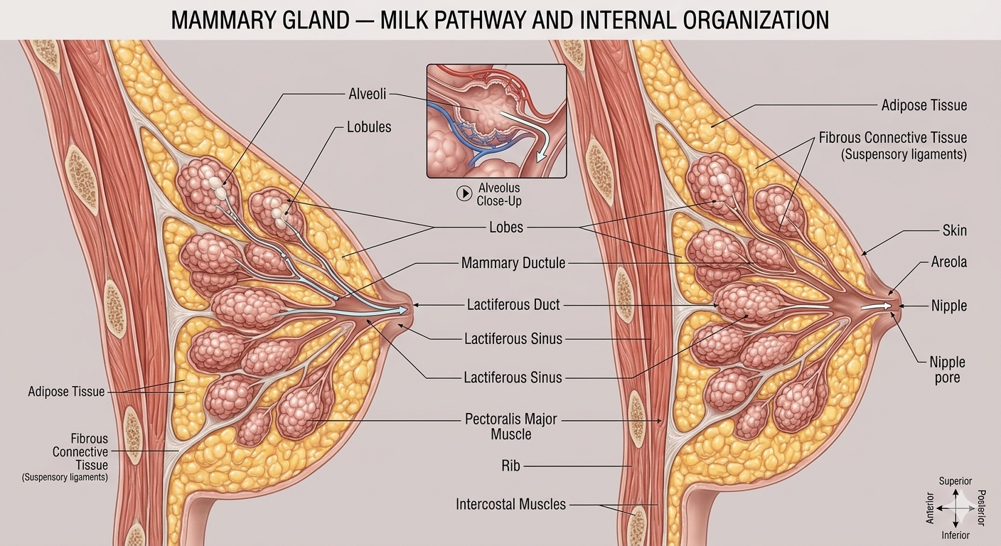
Each breast contains glandular tissue divided into 15–20 mammary lobes with alveoli. Alveoli secrete milk and have lumens where milk is stored. Alveoli open into mammary tubules; tubules join to form mammary ducts; several ducts form a mammary ampulla connected to a lactiferous duct that opens at the nipple.
Formation of sperm in males and the mature female gamete through oogenesis.
The primary sex organs — testis in males and ovaries in females — produce gametes by gametogenesis. In the testis, sperm formation is called spermatogenesis; in the ovary, formation of the mature female gamete is called oogenesis.
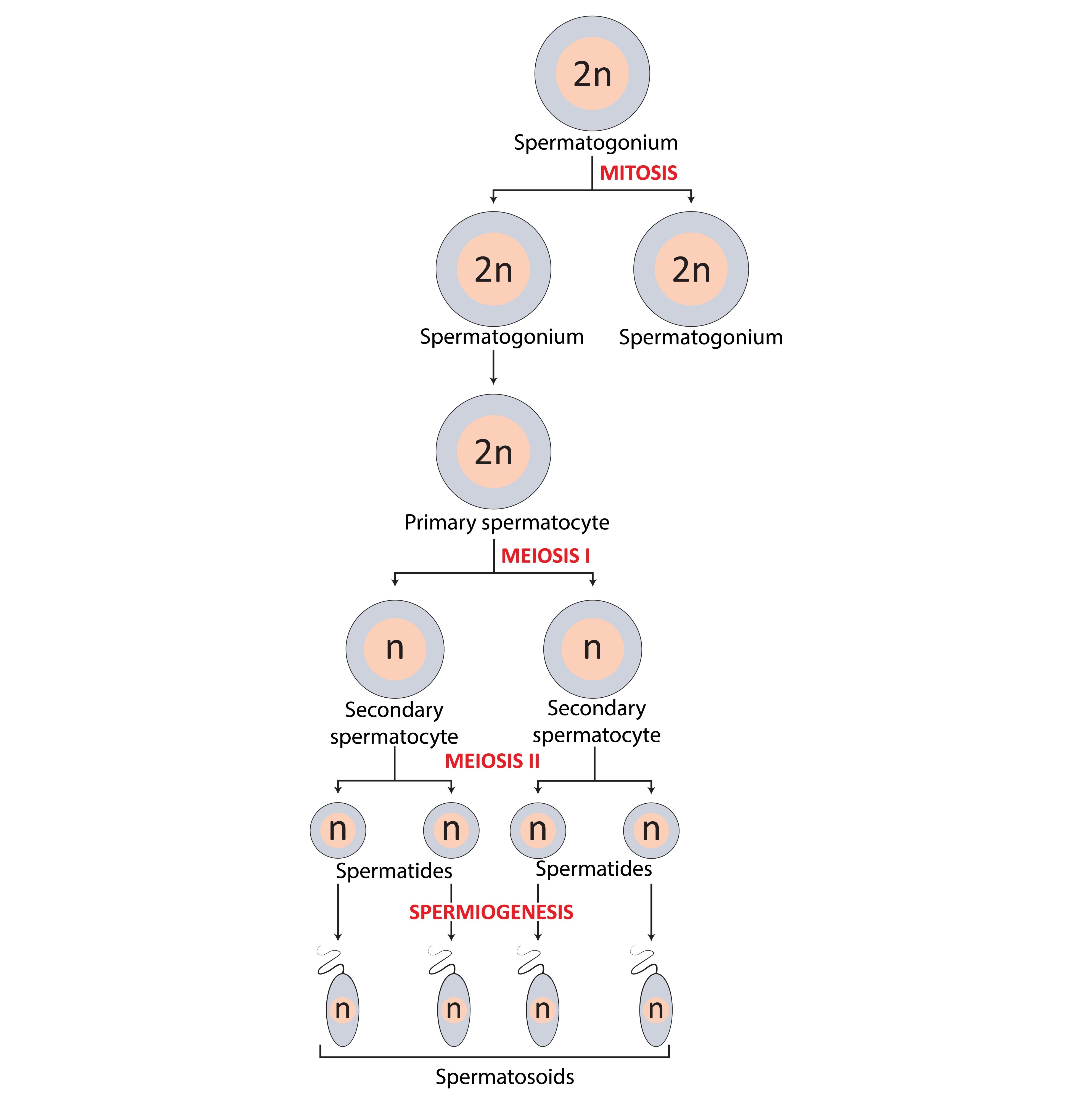
Spermatogonia present on the inside wall of seminiferous tubules multiply by mitotic division. They are diploid and contain 46 chromosomes. Primary spermatocytes undergo meiosis I to form two equal haploid secondary spermatocytes, each with 23 chromosomes. Secondary spermatocytes undergo meiosis II to form four equal haploid spermatids. Spermatids become spermatozoa by spermiogenesis, and mature sperm are released from the seminiferous tubules by spermiation.
Spermatogenesis begins at puberty with increased secretion of GnRH. GnRH stimulates the anterior pituitary to secrete LH and FSH. LH acts on Leydig cells and stimulates androgen synthesis and secretion; androgens in turn stimulate spermatogenesis. FSH acts on Sertoli cells and stimulates secretion of factors that support spermiogenesis.
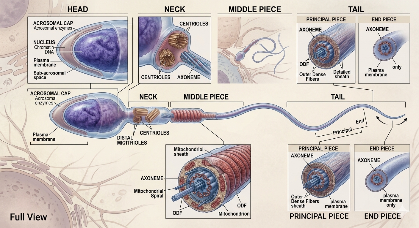
A sperm is composed of head, neck, middle piece and tail. The plasma membrane surrounds the cell. The head contains an elongated haploid nucleus; its anterior region is covered by the cap-like acrosome. The acrosome contains enzymes that help fertilisation. The middle piece contains numerous mitochondria that produce energy for tail movement.
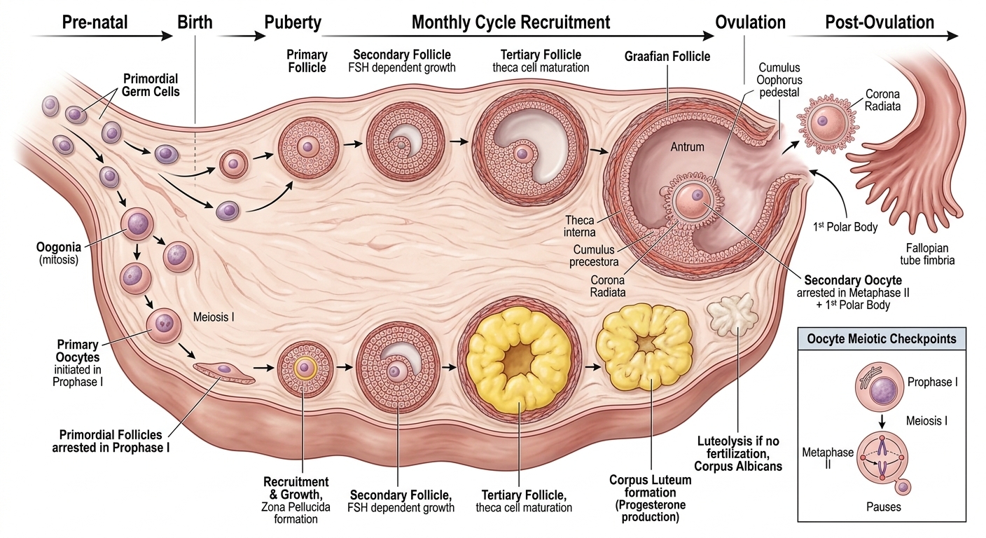
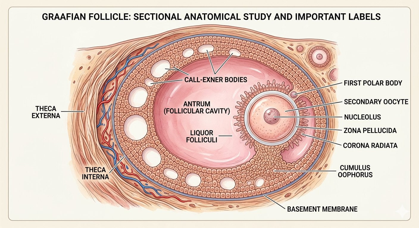
Oogenesis begins during embryonic development. Oogonia form within the fetal ovary; after birth no more oogonia are formed or added. These cells enter prophase I and become primary oocytes. Each primary oocyte becomes surrounded by granulosa cells to form a primary follicle. Many follicles degenerate between birth and puberty, leaving about 60,000–80,000 primary follicles in each ovary at puberty. Primary follicles develop into secondary follicles, then tertiary follicles characterised by a fluid-filled antrum. The theca becomes organised into theca interna and theca externa. The primary oocyte completes its first meiotic division unequally to form a large haploid secondary oocyte and a tiny first polar body. The secondary oocyte forms a zona pellucida, the follicle becomes the mature Graafian follicle, and rupture releases the secondary oocyte by ovulation.
Ovarian and uterine changes controlled by pituitary and ovarian hormones.
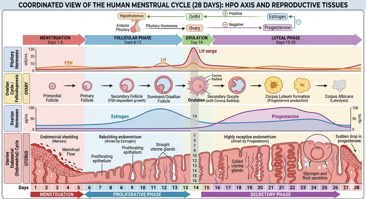
The reproductive cycle in female primates is called the menstrual cycle. In humans, the cycle repeats at an average interval of about 28/29 days. Menarche is the first menstruation at puberty; menopause is cessation of menstrual cycles around 50 years of age.
The cycle begins with menstrual flow and this lasts 3–5 days. The flow results from breakdown of the endometrial lining and its blood vessels, producing fluid that exits through the vagina. Menstruation occurs when the released ovum is not fertilised.
Primary follicles grow toward a mature Graafian follicle, while the endometrium regenerates through proliferation. LH and FSH gradually increase and stimulate follicular development and estrogen secretion by growing follicles.
LH and FSH reach peak levels around the middle of the cycle, about the 14th day. Rapid secretion of LH produces the LH surge, which induces rupture of the Graafian follicle and release of the ovum.
The remaining parts of the Graafian follicle form the corpus luteum, which secretes large amounts of progesterone. Progesterone is essential for maintenance of the endometrium, which is necessary for implantation and other events of pregnancy. If fertilisation does not occur, the corpus luteum degenerates and endometrium disintegrates, beginning a new cycle.
From insemination and sperm transport to zygote, cleavage, blastocyst and implantation.
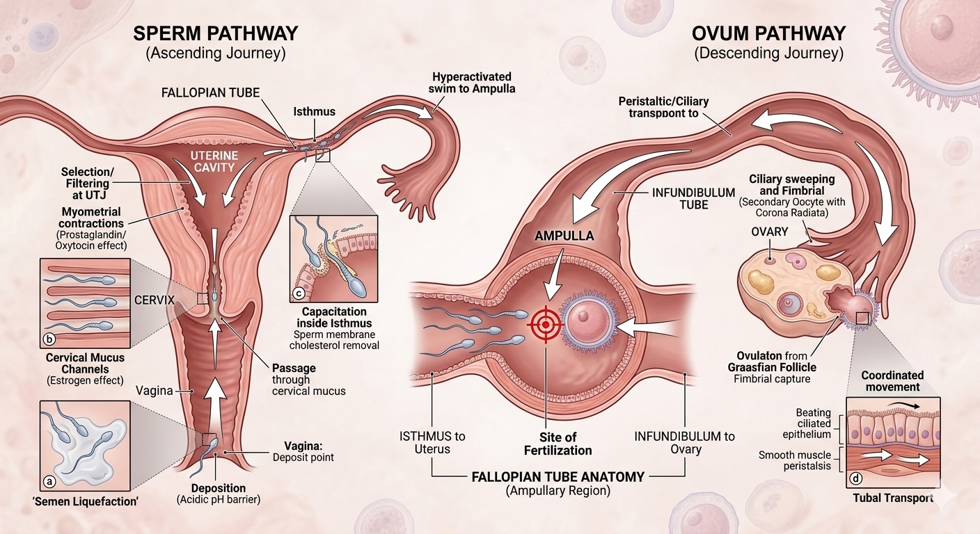
During coitus, semen is released into the vagina (insemination). Motile sperm pass through the cervix, enter the uterus and reach the ampullary region of the fallopian tube. The ovum is also transported to the ampullary region, where fertilisation takes place if sperm and ovum arrive there simultaneously.
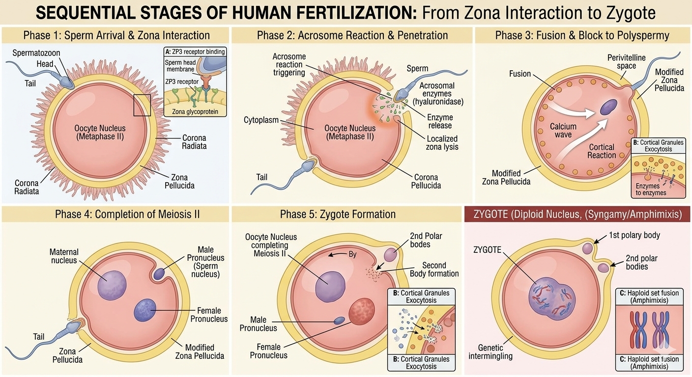
A sperm comes in contact with the zona pellucida of the ovum and induces changes that block entry of additional sperms. Acrosome secretions help the sperm enter through the zona pellucida and plasma membrane. This triggers completion of the second meiotic division of the secondary oocyte, producing a second polar body and a haploid ovum (ootid). The haploid sperm nucleus and ovum nucleus then fuse to form a diploid zygote.
The source explains that human females have XX and ova carry X; male sperms may carry X or Y. Therefore, a zygote may be XX or XY depending on whether an X-bearing or Y-bearing sperm fertilises the ovum. The source explicitly notes that this is why scientifically the sex of the baby is determined by the sperm contribution rather than the mother.
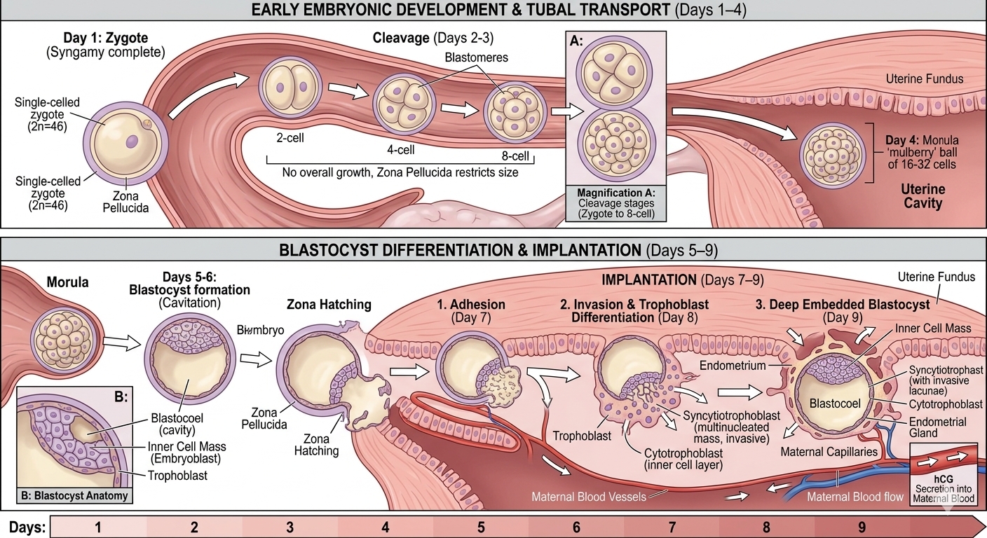
The zygote undergoes mitotic division as it moves through the isthmus toward the uterus. Cleavage forms 2, 4, 8 and 16 blastomeres. An embryo with 8–16 blastomeres is called a morula.
Placenta, pregnancy hormones, germ layers and developmental milestones.
After implantation, finger-like projections called chorionic villi appear on the trophoblast. These are surrounded by uterine tissue and maternal blood. Chorionic villi and uterine tissue become interdigitated and jointly form the placenta, a structural and functional unit between the developing embryo/foetus and maternal body.
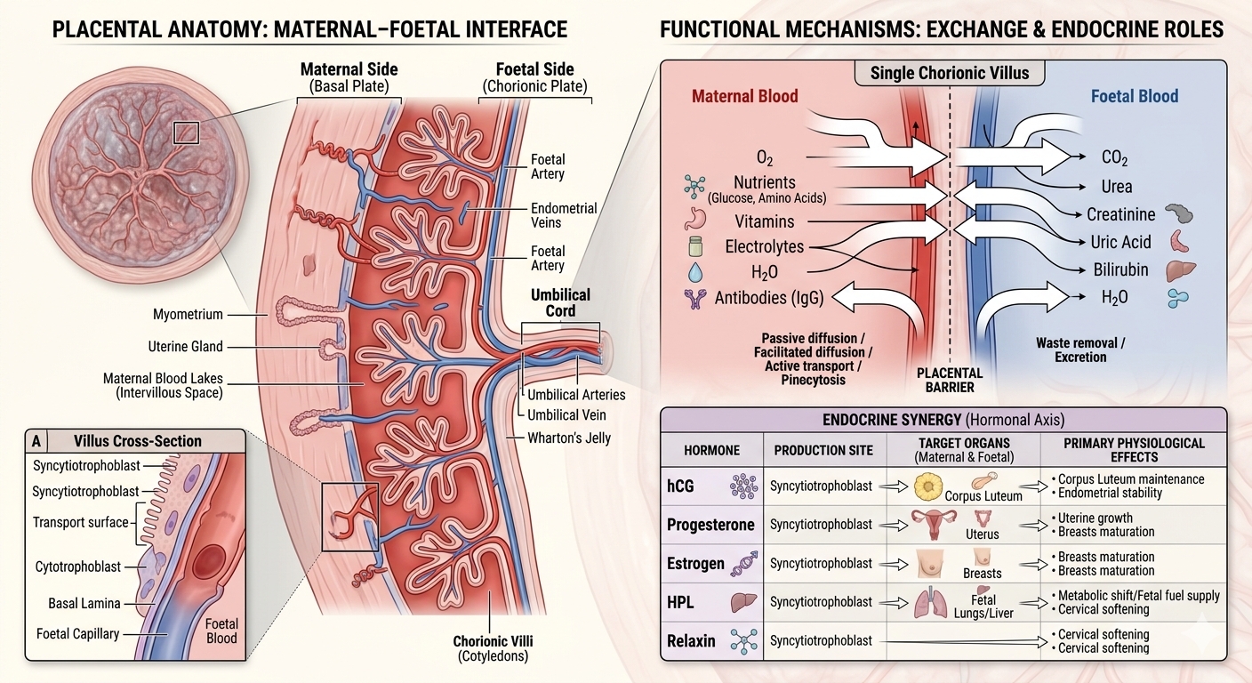
The placenta produces hCG, hPL, estrogens and progestogens. In later pregnancy, relaxin is also secreted by the ovary. The source also notes increased maternal levels of estrogens, progestogens, cortisol, prolactin and thyroxine during pregnancy, supporting foetal growth, maternal metabolic changes and maintenance of pregnancy.
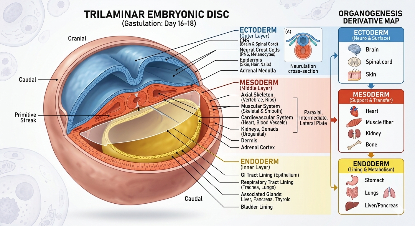
Immediately after implantation, the inner cell mass differentiates into an outer ectoderm and an inner endoderm, with mesoderm appearing between them. These three layers give rise to all tissues and organs in adults. The source also notes that the inner cell mass contains stem cells with the potency to give rise to tissues and organs.
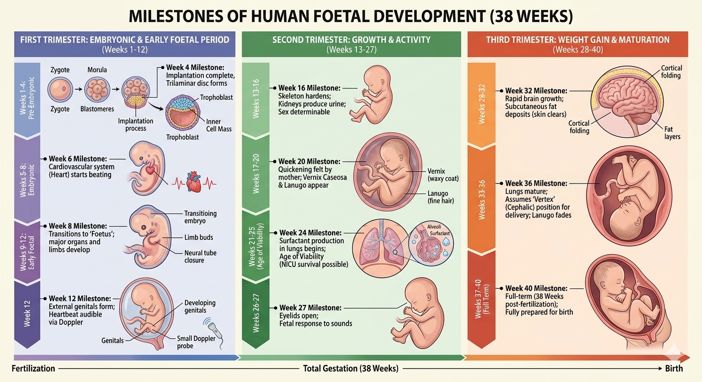
| Time | Source-described milestone |
|---|---|
| 1 month | Embryo’s heart is formed. |
| 2 months | Limbs and digits develop. |
| 12 weeks | Most major organ systems are formed; limbs and external genital organs are well developed. |
| 5 months | First foetal movements and appearance of hair on the head are usually observed. |
| About 24 weeks | Fine hair covers the body; eyelids separate and eyelashes form. |
| 9 months | Foetus is fully developed and ready for delivery. |
Birth through neuroendocrine feedback, followed by milk production and newborn feeding.
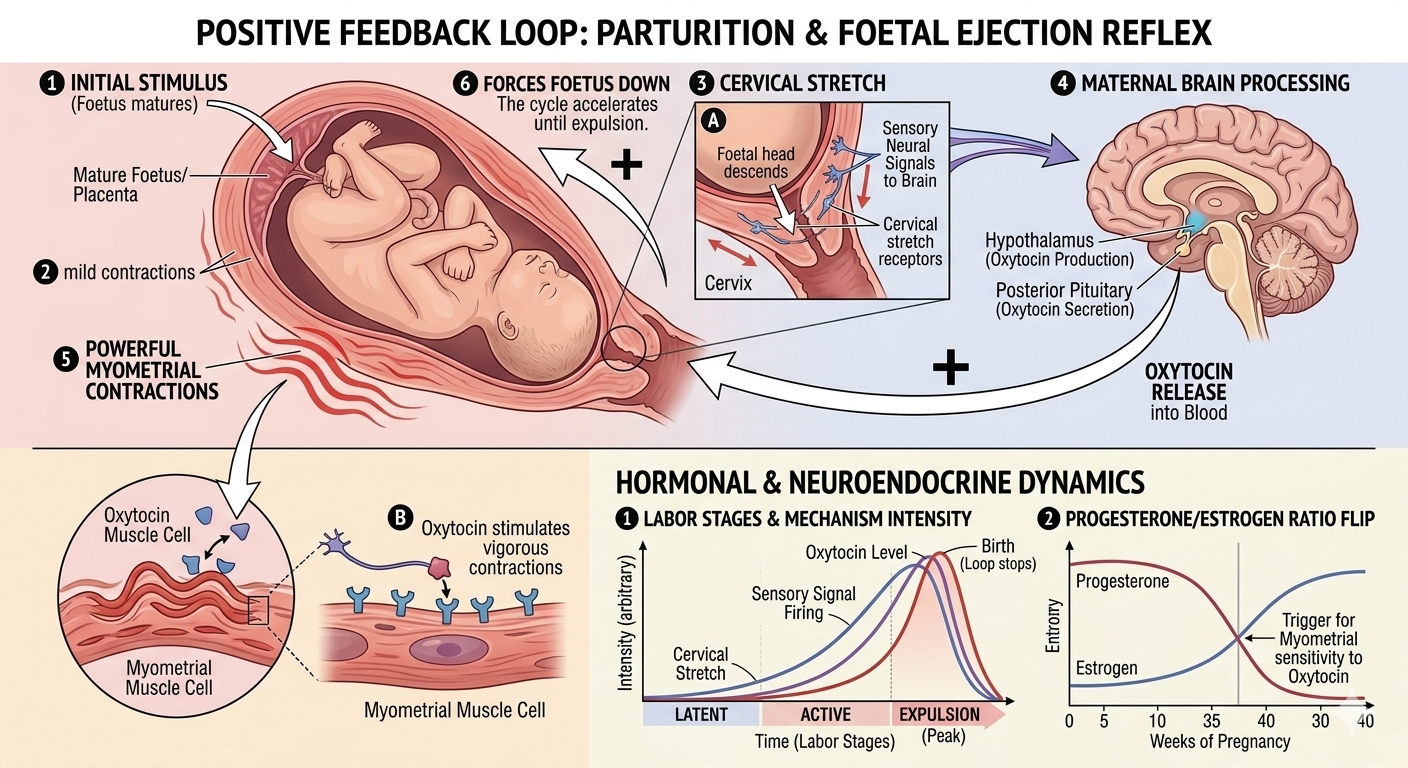
The average duration of human pregnancy is about nine months, called the gestation period. Vigorous uterine contractions at the end of pregnancy cause expulsion of the foetus. This childbirth process is called parturition.
Signals from the fully developed foetus and placenta induce mild uterine contractions called the foetal ejection reflex. This triggers oxytocin release from the maternal pituitary. Oxytocin acts on uterine muscle and strengthens contractions. Stronger contractions stimulate further oxytocin secretion, creating a positive feedback loop that continues until the baby is expelled through the birth canal. The placenta is expelled soon after delivery.
Mammary glands differentiate during pregnancy and begin producing milk toward the end of pregnancy. This process is called lactation. Milk produced during the initial few days is called colostrum and contains several antibodies that are important for developing resistance in the newborn.
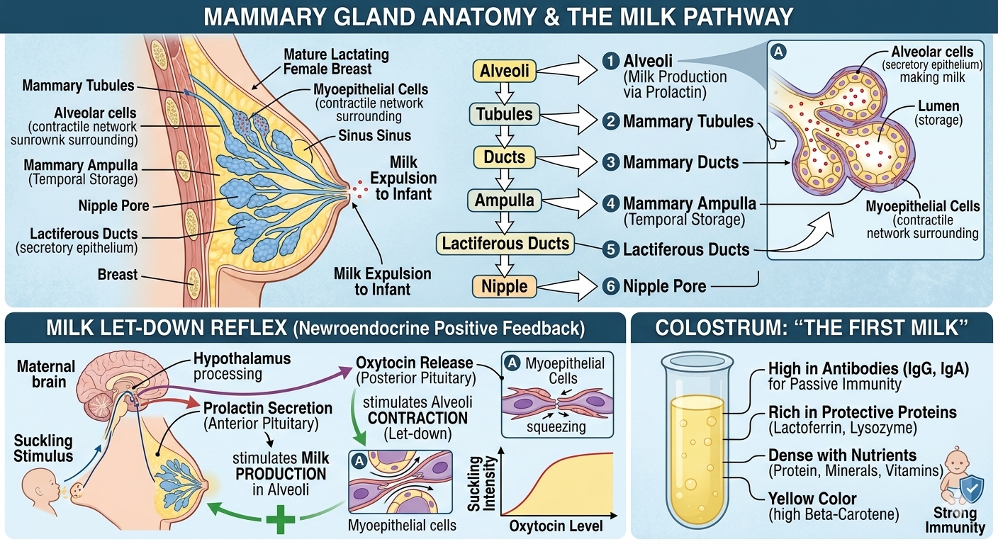
One screen for the chapter’s most important sequences and facts.
Exercises represented from the supplied chapter.
Write the functions of: corpus luteum, endometrium, acrosome, sperm tail, fimbriae.
Chapter-based answers prepared directly from the supplied NCERT chapter. Use the answers after attempting the questions yourself.
First solve the exercise without looking below. Then compare your response with the chapter-based answer. Long answers are written in an exam-friendly form while keeping the source chapter's terminology and sequence.
| Structure | Function |
|---|---|
| Corpus luteum | Secretes large amounts of progesterone, which is essential for maintenance of the endometrium. |
| Endometrium | Inner glandular layer of the uterus; it undergoes cyclical changes and provides the uterine lining important for implantation and pregnancy maintenance. |
| Acrosome | Cap-like structure on the sperm head containing enzymes that help the sperm enter the ovum during fertilisation. |
| Sperm tail | Facilitates sperm motility so that the sperm can move through the female genital tract. |
| Fimbriae | Finger-like projections of the infundibulum that help in collection of the ovum after ovulation. |
Select your answers and click "Check Answers"